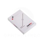
MOPTIM DEA-Dry Eye Analyzer


Brand: Moptim
MOPTIM DEA-Dry Eye Analyzer
MOPTIM Dry Eye Analyzer (DEA)
Specification:
IMAGE AND VIDEO ACQUISITION
- Image Resolution: 8,000,000 pixels
- Image Dimensions: 3864 x 2218 pixels JPEG
- Acquisition Mode: Multi-shot photos, video
- Focus and Exposure: Manual and automatic
- Covering Area: Maximum 8 mm
- Camera: Colored, sensitive to infrared
- Light Source: Red, blue and white LED
GENERAL INFORMATION
- Working Distance: 5 mm – 50 mm
- Ports: USB 3.0
- Power Supply: 5 V
- Dimension: 170mm (H) x 54mm (W) x 64~110mm (L)
- Weight: 427g (including main body, 4 lenses and 1 wireless camera shutter)
- Accessories: Standard: wireless camera shutter, lenses, briefcase, lens case, slit lamp adaptor Optional: complete holder, instrument table
SOFTWARE AND DATA MANAGEMENT
- Operating System: Windows 10 64 bit
- System Requirement: Intel core i3, RAM 8GB, hard disk 200G, screen resolution: 1920*1080
- Exams: DEQ-5, fluorescein staining, NIBUT, FBUT, interferometry, auto meibomian gland evaluation, auto tear meniscus height, auto redness, eyelid margin imaging, anterior imaging
The DEA by MOPTIM is an innovative dry eye diagnostic system that performs complete examinations for dry age with outstanding imaging capability. The compact design makes it easy to integrate into any practice seamlessly
MOPTIM DEA-Dry Eye Analyzer
Overview:
- Comprehensive 10-in-1 exam capability for ocular surface evaluation.
- 8 mp high resolution camera and outstanding optical design bring high level of detail and clarity.
- Effortless precision with automated functions: auto NIBUT, tear meniscus height, meibomian gland analysis, and redness detection.
- Intuitive software that guides you with ease and allows for personalized exam protocols, tailored to your needs.
- Compact design for easy installation on any slit lamp or handheld use.
- Auto meibography
Evaluate the meibomian glands with red light, the software provides automatic evaluation of loss area. - Non-invasive breakup time
Automatically analyze break-up area, first and average break-up time for tear stability evaluation. - Interferometry
Record a video of blinking process to observe the surface reflection pattern and dynamics of the tear film. - Tear meniscus height
Automatically evaluate tear meniscus height that is observed on the eyelid margins. Up to 5 measurement points can be taken. - Fluorescein Staining
Evaluate the areas of damage on the ocular surface after application of the fluorescein dye. Compare your images with grading scales incorporated in the software. - Auto Redness
Eye redness could be one of the symptoms of dry eye disease. Automatically compare your images with grading scales incorporated in the software. - Eyelid margin imaging
MGD can cause the glands to become blocked, impacted, and infected. Capture high resolution under white LED illumination, and compare your images with grading scales included in the software. - Color-coded reports for quick dry eye insights
The DEA Dry Eye Analyzer’s easy-to-read report template with color coding transforms data interpretation into an intuitive experience.